About Eye Tracking
The role of eye movements in dyslexia has been debated for many years. It is known from studies that dyslexics have an increase in the duration of fixations (the pauses that we make when reading) and that they make more backwards eye movements (called “regressions”). These are usually explained however as a manifestation of poor comprehension rather than as a primary abnormality of the eye movements themselves. Historically, this was demonstrated by a number of studies which showed little or no difference in eye movements between dyslexics and non-dyslexics.1,2,3 Although this has become the popular view, it is not supported using currently accepted methods for evaluating eye movements.
Pursuits  Pursuit eye movements (eg. follow the ball on the end of a stick) are not often considered in the scientific literature on dyslexia since we do not use this type of eye movement to read a book. Nevertheless, large studies show a strong correlation between the quality of pursuit eye movements and reading difficulties in young children4,5,6 – an observation commonly made by optometrists and teachers. A possible explanation for this is the more recent understanding that pursuits and saccades (although distinctly different types of eye movements) are controlled by similar networks of cortical and sub-cortical regions in the brain, and in some cases even share the same neurons!7 Since the development of pursuit eye movements reach adult maturity earlier than saccades their predictive value based on observation alone rapidly diminishes beyond the early school years. The pictures below show how pursuit eye movements can look for a non-dyslexic child (blue) compared to a dyslexic child (red) when using the latest eye tracking technology.8
Saccades Saccadic eye movements are a more rapid type of eye movement and comprise of various subtypes including: voluntary saccades, corrective saccades, anticipatory saccades, reflex saccades and express saccades. Voluntary saccades, referred to by researchers as “anti-saccades” (as they are measured by asking the subject to look in the opposite direction of the test stimulus) are the type of saccades employed for reading text and include corrective saccades which are used when correcting saccade errors.
Voluntary Saccades Voluntary saccades are so named because they are controlled by the frontal and prefrontal structures in the brain associated with conscious decision making.9,10
Reflex Saccades By contrast, reflex saccades are controlled by the mid-brain region (eg. the Superior Colliculus) and are a response to rapidly moving stimuli in our peripheral vision (eg. a cricket ball coming from the side).
Fixation Fixations are the regular pauses that we make when we are reading (see diagram below or click on tracker for video demonstration). It is during each fixation period that visual information is processed and used for higher order processing in the brain. Fixation is an active process, and in order to make any kind of saccadic eye movement (voluntary or reflexive) requires suppression (ie. switching off) of the fixation mechanism.
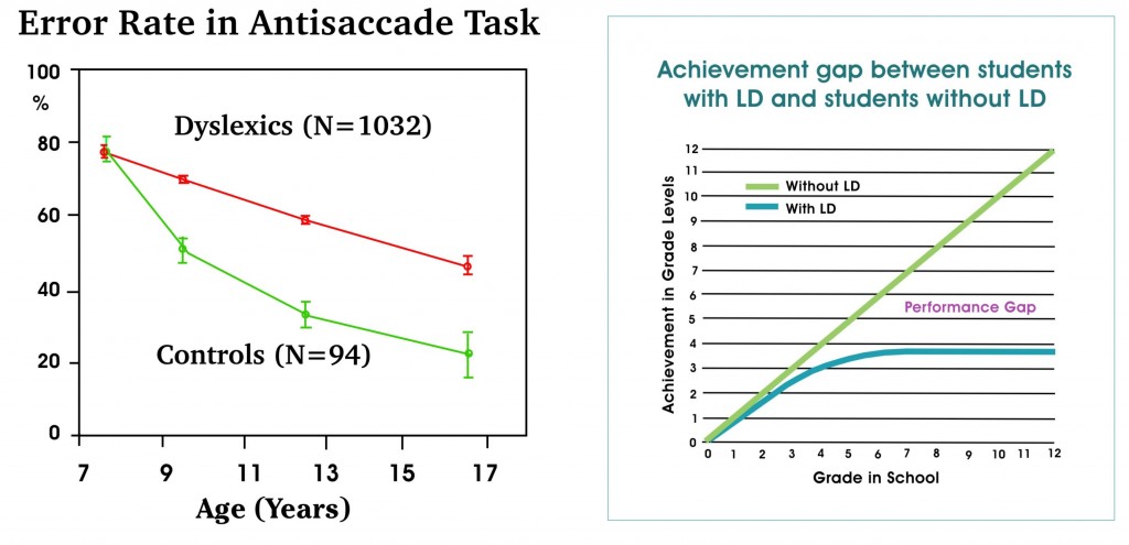
It is argued that in order to determine if an ocular motor abnormality is associated with a specific learning disability, a large group must be assessed with objective eye movement recordings and an age matched control group performing academically well must be studied in exactly the same fashion.11 Such a study was done over 10 years ago involving around 1000 dyslexic children ages 7 to 17 years of age, utilizing a task that is independent of language12. Their findings show firstly a long developmental period for saccades to reach adult maturity in the normal population; up to about 17 years of age in the case of voluntary saccades and around 15 years of age in the case of reflex saccades. Secondly, a significant difference between dyslexics and non-dyslexics in the reaction time of reflex saccades is found for younger children (less than 9 years of age), and for older children (over 13 years of age) when making voluntary saccades. Thirdly, and most importantly, there is a significant difference in the control of voluntary eye movements between dyslexics and normal readers after the age of 7 (as shown by the higher error rate on the graph below) which continues to become greater with age relative to peers, a finding that has been confirmed by another study involving students ages 8 to 16 years of age.13 By around 20 years of age 80% of dyslexics exhibit a problem with the control of their voluntary saccades. It is interesting comparing a graph that shows how the eye tracking deficit changes with age with a graph that shows how achievement in a child with a learning disability changes with age (National Center of Learning Disabilities, 2011). In both cases the performance gap gets worse as the child gets older.
Interestingly, similar problems with saccadic eye movement control have also been reported in children with ADD14,15, Aspergers16, mild closed head injury17,18 and a range of other neurological conditions not discussed in here. It is apparent that voluntary saccades are a very sensitive marker of frontal lobe dysfunction – even in the absence of other neurological signs.
The reason for the historical lack of significance in eye movements between dyslexics and their normal reading peers, can largely be attributable to the scientific methodology used. For example, until recently nearly all of the reported studies relating to saccadic eye movements in dyslexia, are limited to saccadic latencies (ie. reaction times) or amplitudes and do not investigate the control of voluntary saccades as measured by the number of errors made on an anti-saccade task. In the case of saccadic latencies, most studies involve children 9 to 13 years of age and we know (as discussed above) that differences are only found in saccadic latencies outside of these age ranges!
Finally, there is evidence to show that dyslexics have problems with both monocular and binocular fixation stability under 12 years of age.19,20,21,22 The idea of a problem with binocular fixation has been looked at by Professor John Stein of Oxford University who describes this condition as an “eye wobble” thought to be caused by a deficit in the magnocellular visual pathways23.
Training Eye Movements Once it has been established that a child has faulty eye movements it would be remiss not to consider whether these problems might be treatable, particularly when one considers that reading is such a strongly visual task. Not surprisingly, various treatments that usually involve some kind of regular eye movement training have been offered by optometrists for many years and are advocated by the American Optometry Association.24,25 Not everyone agrees however that the training of eye movements is scientifically supported as an efficacious treatment for dyslexia.26,27 The remainder of this article will consider the evidence that supports the training of eye movements, since there is little evidence to show that they cannot be trained.
There are relatively few studies in the mainstream literature to show that pursuit eye movements can be improved with training, and such studies are probably more likely to be found in clinical reports and journals.28,29,30 One such example is the report by the Reading & University Laboratory of Physiology at the Royal Berks Hospital, Oxford, in which an audit of treatments involving some 300 dyslexic children in their Learning Difficulties Clinic, showed that pursuits, saccades and convergence are all significantly improved with training leading to improvements in reading of around 2 months/per month; a result that compares favorably with traditional approaches.31
By comparison, there are numerous clinical and research studies demonstrating that saccadic eye movements can be trained to – if not close to – age normal levels,32,33,34,35,36 and that training has a positive effect on academic outcomes, including children with developmental dyslexia.37,38,39,40,41,42 One such article, by the Optomotor Laboratory Brain Research Group in Germany, shows that voluntary saccadic eye movements can be improved by over ten times their natural rate of development following 10 minutes of daily training, using a small hand held device for a period of 3 to 8 weeks.32 This cannot be attributable to a placebo effect since the same device used for training reflex saccades has no such affect.
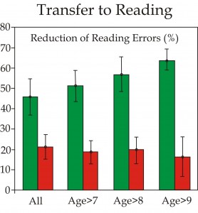
Such a treatment can result in a 50% average reduction in reading errors compared to only a 20% reduction with controls (see graph). Both groups received 6 weeks of formal reading instruction.37 Similarly, a randomized placebo controlled study by Stein showed that binocular stability can be improved in dyslexics with monocular occlusion therapy resulting in significant reading gains for dyslexic children.43 Although the use of the Dunlop Test to determine binocular stability has been questioned44, similar findings have been reported by other researchers. One such finding showed that alternate monocular occlusion can reduce binocular fixation instability by around 55%.45-48
Summary  There now exists a growing body of evidence to demonstrate that eye movement dysfunction, both in terms of voluntary saccadic control, binocular fixation stability and also saccadic latencies in the younger and older student populations, significantly contributes towards the dysfunction of developmental dyslexia. Such a finding is not surprising when one considers the kinds of problems frequently observed in dyslexics including skipping letters or words, losing place and difficulty with visual search activities. These problems cannot be solely attributed to a primary language disorder as has previously been held to be the case. Indeed had this been understood much earlier the intervention for dyslexia would probably look quite different today given the dominant role of vision in reading.
The oculomotor dysfunctions observed in dyslexia may possibly be explained in terms of a magnocellular (M-pathway) deficit due to the high number of magnocellular projections into the brain structures responsible for the control of voluntary eye movements. Furthermore, it has previously been established that the magnocellular pathways of dyslexics have both anatomical and functional abnormalities in the lateral geniculate nucleus, extrastriate visual cortex Area MT/V5 and parietal regions.46,47,48 My purpose however, is not to present a neurobiological basis for dyslexia but to demonstrate the importance of diagnosing eye movement disorders in children failing to progress at school despite having received appropriate instruction, especially when a diagnosis of dyslexia has been made. In my experience, these problems can usually be improved to age normal levels with reported gains in academic performance. The interventions for improving eye movements should not be considered as a replacement for traditional therapies, however a combination of non-linguistic treatments such as eye movement training, together with more traditional approaches is the most likely to be effective. This also provides educators with another approach to treatment which could be particularly helpful when traditional therapies are failing. The argument against training on the basis of cost fails to compare this against the cost of delivering existing strategies, not to mention the financial and social cost of failing to remediate the child with a serious learning problem such as dyslexia.


